What Makes Classification Possible
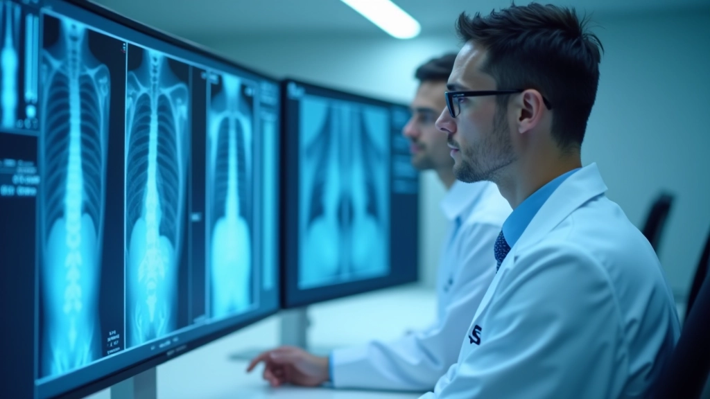
Medical imaging produces enormous amounts of visual data every single day. Radiologists spend hours analyzing subtle differences in pixel patterns — dark spots that might indicate disease, variations in tissue density, structural changes that signal problems. That's where classification systems come in. They're not magic. They're pattern recognition tools trained on thousands of real images.
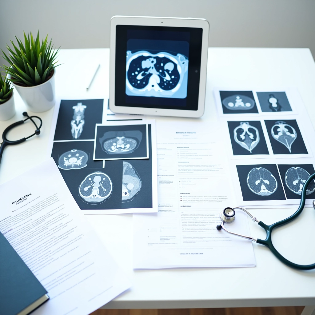
A classification system works like this: you feed it labeled examples. A chest X-ray marked "normal" and another marked "pneumonia." Then another marked "tuberculosis." The system learns to spot the visual differences. Over time, with enough examples, it develops an understanding of what each condition looks like on an image. When you show it a new X-ray it's never seen before, it can make a prediction based on what it learned.
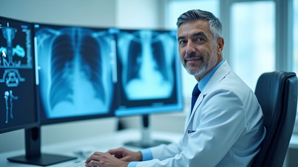
Here's the important part — it's not infallible. It's a tool. A good one when used correctly, but one that works best alongside human expertise. In Edmonton hospitals and clinics, radiologists aren't being replaced. They're getting a second set of eyes that works faster and never gets tired.
Key insight: Classification systems excel at consistency. They evaluate every image by the same criteria, every single time. A radiologist might miss something on their 200th case of the day. The AI doesn't have that problem.
How Neural Networks Learn Patterns
The technology behind this is called a convolutional neural network, or CNN. Don't let the name intimidate you. Think of it as a system with layers. Each layer looks for different things.
The first layers spot simple stuff — edges, corners, brightness changes. It's like your brain's visual cortex recognizing basic shapes before understanding what they mean. The next layers combine those simple patterns into more complex ones. Circles. Lines. Structures. Deeper layers recognize actual anatomical features — ribs, lung tissue, the heart outline.
By the final layers, the system's learned to recognize what actually matters. A nodule that looks suspicious. A fracture line. Fluid accumulation where there shouldn't be any. It's learned these patterns by looking at thousands of labeled examples and gradually adjusting its internal weights to predict correctly.
The Training Data Question
Here's where things get practical. A classification system is only as good as the data it learns from. If you train it on 5,000 chest X-rays from one hospital with one type of scanner, it might struggle when it sees images from a different facility with different equipment. The pixel patterns look subtly different.
Quality matters more than quantity. You're better off with 2,000 carefully labeled, diverse images than 20,000 low-quality ones with errors in the labels. When a radiologist marks an image incorrectly — says it's normal when it actually shows pathology — the system learns the wrong thing. Garbage in, garbage out, as they say in tech.
Modern implementations focus on what's called "domain adaptation." That means you take a system trained on a massive general dataset and then fine-tune it using your hospital's specific images. This approach works better because it starts with broad pattern knowledge, then learns the quirks of your particular scanners and patient populations.