MediScan AI Logo MediScan AI Contact Us

Understanding Medical Imaging Classification Systems

How AI algorithms learn to identify patterns in X-rays, CT scans, and MRIs — the fundamentals that make automated diagnostics possible in clinical settings.

July 2026 12 min read Intermediate
MediScan AI Editorial Team profile photo

Author

MediScan AI Editorial Team

Editorial Team

Written by the MediScan AI Editorial Team, focused on practical, honest guidance for healthcare professionals implementing AI in diagnostic workflows.

What Makes Classification Possible

Medical imaging produces enormous amounts of visual data every single day. Radiologists spend hours analyzing subtle differences in pixel patterns — dark spots that might indicate disease, variations in tissue density, structural changes that signal problems. That's where classification systems come in. They're not magic. They're pattern recognition tools trained on thousands of real images.

A classification system works like this: you feed it labeled examples. A chest X-ray marked "normal" and another marked "pneumonia." Then another marked "tuberculosis." The system learns to spot the visual differences. Over time, with enough examples, it develops an understanding of what each condition looks like on an image. When you show it a new X-ray it's never seen before, it can make a prediction based on what it learned.

Here's the important part — it's not infallible. It's a tool. A good one when used correctly, but one that works best alongside human expertise. In Edmonton hospitals and clinics, radiologists aren't being replaced. They're getting a second set of eyes that works faster and never gets tired.

Key insight: Classification systems excel at consistency. They evaluate every image by the same criteria, every single time. A radiologist might miss something on their 200th case of the day. The AI doesn't have that problem.

How Neural Networks Learn Patterns

The technology behind this is called a convolutional neural network, or CNN. Don't let the name intimidate you. Think of it as a system with layers. Each layer looks for different things.

The first layers spot simple stuff — edges, corners, brightness changes. It's like your brain's visual cortex recognizing basic shapes before understanding what they mean. The next layers combine those simple patterns into more complex ones. Circles. Lines. Structures. Deeper layers recognize actual anatomical features — ribs, lung tissue, the heart outline.

By the final layers, the system's learned to recognize what actually matters. A nodule that looks suspicious. A fracture line. Fluid accumulation where there shouldn't be any. It's learned these patterns by looking at thousands of labeled examples and gradually adjusting its internal weights to predict correctly.
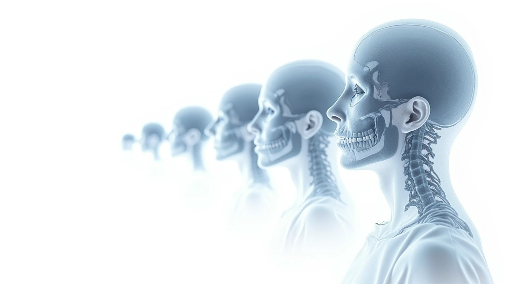
Medical imaging classification neural network visualization showing layers of feature extraction from pixel data to anatomical diagnosis

The Training Data Question

Here's where things get practical. A classification system is only as good as the data it learns from. If you train it on 5,000 chest X-rays from one hospital with one type of scanner, it might struggle when it sees images from a different facility with different equipment. The pixel patterns look subtly different.

Quality matters more than quantity. You're better off with 2,000 carefully labeled, diverse images than 20,000 low-quality ones with errors in the labels. When a radiologist marks an image incorrectly — says it's normal when it actually shows pathology — the system learns the wrong thing. Garbage in, garbage out, as they say in tech.

Modern implementations focus on what's called "domain adaptation." That means you take a system trained on a massive general dataset and then fine-tune it using your hospital's specific images. This approach works better because it starts with broad pattern knowledge, then learns the quirks of your particular scanners and patient populations.
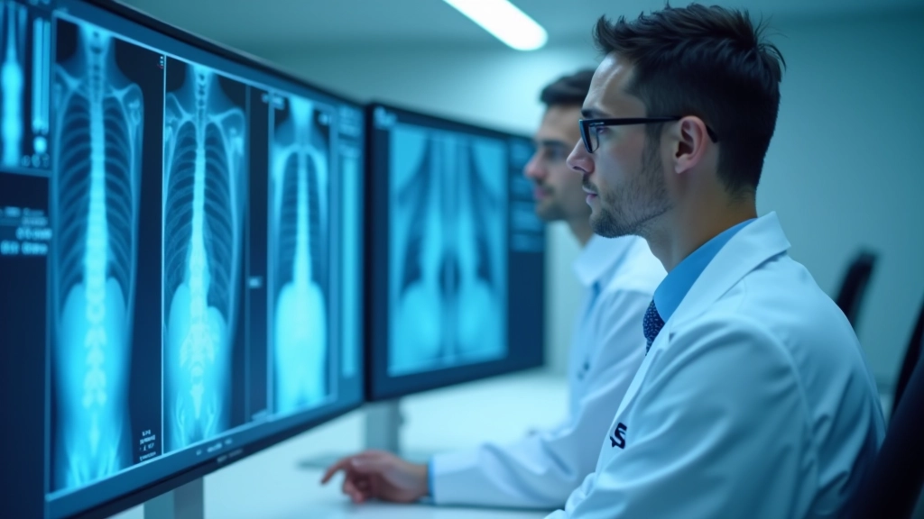

Healthcare professionals reviewing medical imaging data on monitors in a modern diagnostic center

Important note: This article is informational only and is not medical advice. Please consult a qualified healthcare professional about your individual situation or diagnostic needs.

From Classification to Clinical Workflow

Getting a classification system working in a hospital isn't just about accuracy numbers. It's about integration. A radiologist in Edmonton doesn't want to use three separate tools. They want the AI output integrated into their existing workflow. That means the system needs to flag suspicious findings, provide confidence scores, and work with PACS systems — the software that manages medical images.

The best implementations we've seen use what's called "triage classification." The AI doesn't try to make the final diagnosis. Instead, it prioritizes cases. Urgent findings go to the top of the queue. Clearly normal images get reviewed faster. This approach respects radiologist expertise while using AI's speed to improve patient care.

Implementation also requires validation. Before a system touches a single patient case, it's tested on thousands of images it's never seen. You want to know its sensitivity — can it catch the disease when it's actually there? — and its specificity — does it avoid false alarms? These numbers matter because missing a case is worse than creating extra work with a false positive.

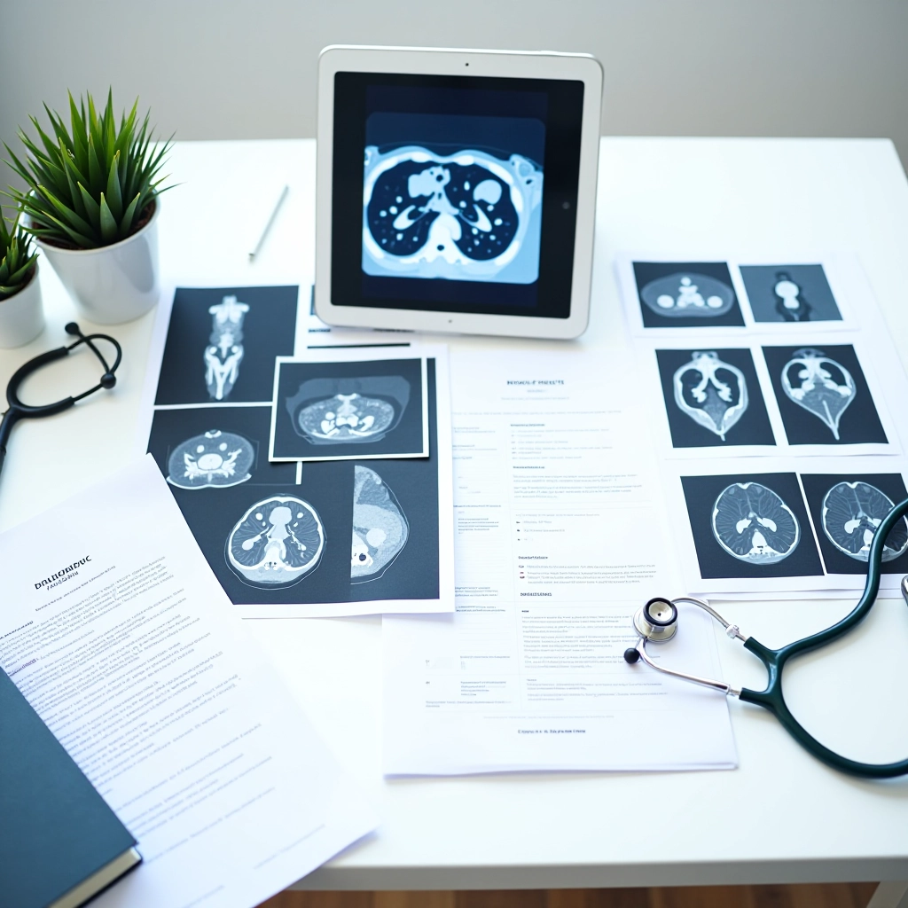
Hospital IT infrastructure with servers and networking equipment in a secure medical data center

Real Limitations Worth Understanding

Classification systems have real constraints. They work best on clear-cut cases. A normal chest looks normal. Pneumonia has recognizable patterns. But medicine isn't always clear-cut. You've got unusual presentations. Patients with multiple conditions. Images from equipment the system wasn't trained on.

They can also develop what's called "bias." If your training data came mostly from certain patient demographics, the system might perform differently on other groups. That's why modern implementations use diverse datasets. Edmonton's healthcare system has committed to this — building classification tools that work across different patient populations, not just on whoever happens to be in the initial training set.

And there's the explainability challenge. Some systems can show you what parts of the image influenced their decision — heatmaps highlighting suspicious regions. Others work like black boxes. You get a prediction but not the reasoning. Clinical settings increasingly demand explainability because radiologists need to understand why the AI flagged something.

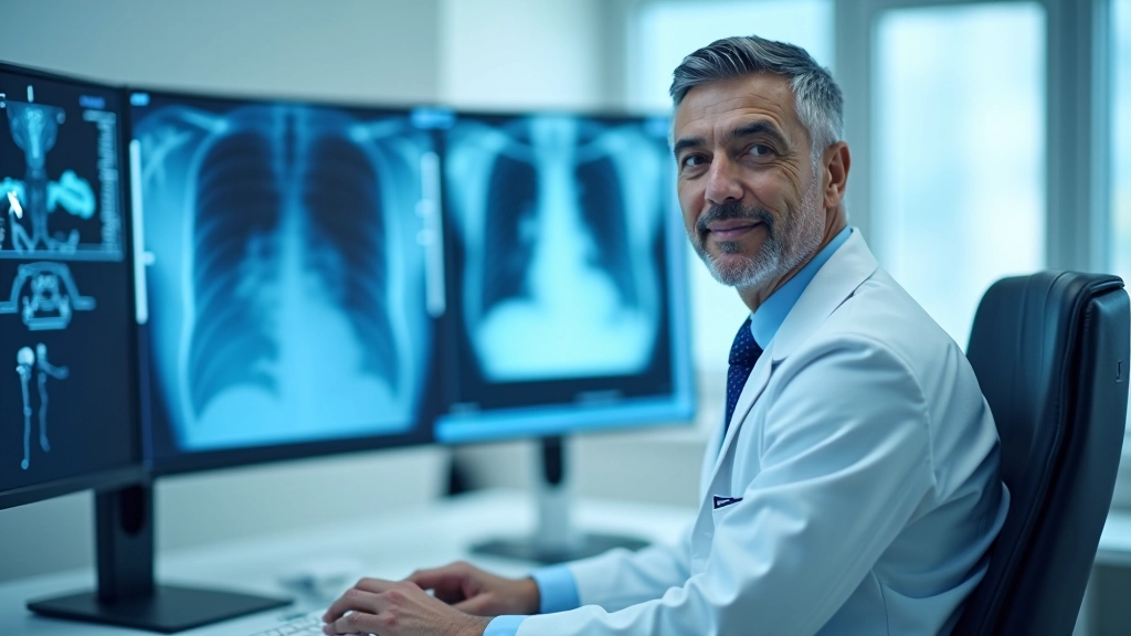
Radiologist at desk with multiple monitors showing medical imaging analysis with AI overlay and diagnostic tools

The Path Forward

Classification systems are getting better at handling edge cases. Newer architectures can learn from smaller datasets. Transfer learning — using knowledge from one task to improve another — is becoming standard practice. And there's growing focus on making these systems work across different imaging equipment and protocols.

For healthcare professionals in Edmonton implementing these tools, the key is understanding that classification isn't about replacing radiologists. It's about extending their capabilities. Faster preliminary screening. Better prioritization of urgent cases. Reduced diagnostic variability. Fewer errors from fatigue.

The systems work best when they're built with input from the people who'll actually use them. Radiologists know what information matters. They understand clinical workflow. When classification systems are designed with that expertise, they integrate smoothly and actually improve patient care instead of just adding complexity.