
Digital Pathology and AI-Assisted Tissue Analysis
How artificial intelligence is transforming microscopy, accelerating diagnoses, and improving accuracy in tissue examination — a practical guide for pathology teams implementing these tools in clinical settings.
By
Editorial Team focused on practical, honest guidance for healthcare professionals implementing AI in diagnostic workflows.
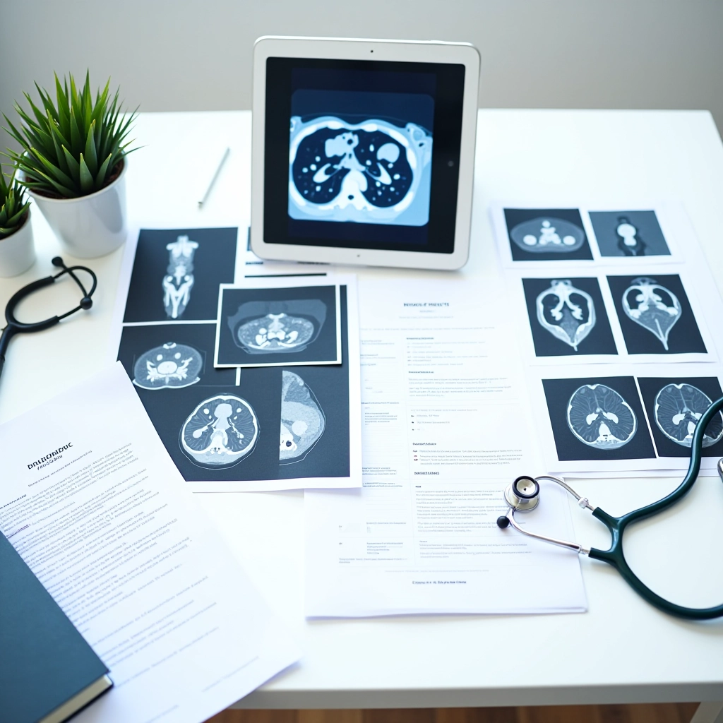
Digital pathology represents one of the most significant shifts in laboratory medicine over the past decade. It's not just about replacing microscopes with screens — it's fundamentally changing how pathologists work, collaborate, and make decisions. When combined with AI-assisted analysis, digital pathology becomes a force multiplier: faster slide scanning, more consistent pattern recognition, and better support for complex diagnostic cases.
But here's the reality: implementing these systems requires careful planning, staff training, and a clear understanding of what AI can and can't do. We've seen teams get excited about the technology and stumble on the practical side. That's what this guide covers — the real implementation story.
What Digital Pathology Actually Does
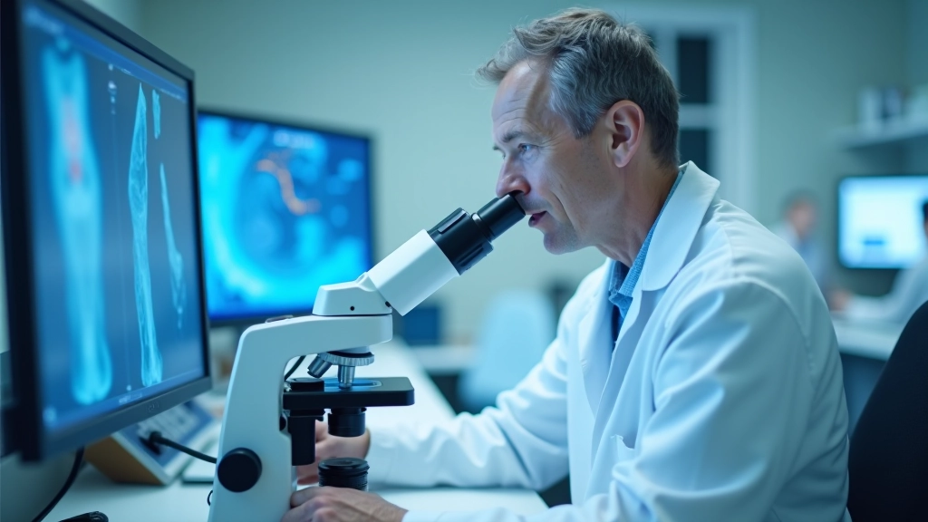
Traditional pathology relies on a pathologist looking at a glass slide under a microscope. Digital pathology scans that slide into a high-resolution image file — typically 40,000 50,000 pixels or larger for a standard slide. You can then view, zoom, and annotate that image on a computer or tablet, anywhere in your facility (or even remotely).
The scanning happens in minutes. A whole-slide image capture takes about 8-12 minutes per slide depending on magnification and tissue type. Once digitized, that image is stored and can be reviewed, shared with specialists, or analyzed by AI algorithms without ever touching the original glass slide again.
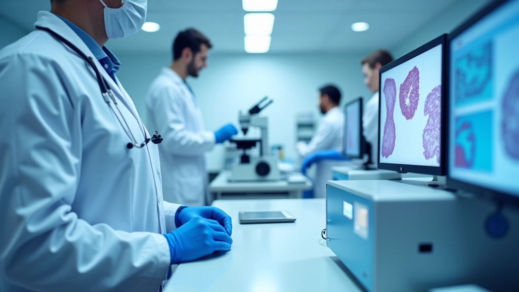
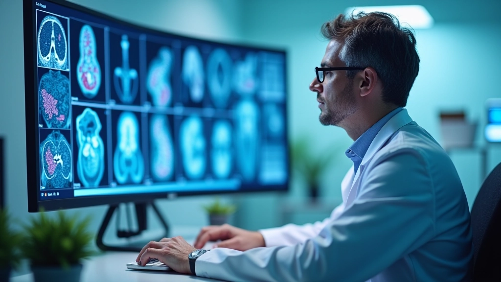
AI enters the picture by analyzing these digital images automatically. Instead of a pathologist scanning every millimeter of a slide looking for abnormalities, AI can pre-screen slides, highlight suspicious regions, count specific cell types, or detect patterns that might take a human hours to identify manually. The pathologist then validates or refines the AI's findings.
Why Pathology Teams Are Adopting This Now
The push toward digital pathology isn't just about technology for technology's sake. Three practical pressures drive adoption right now.
Workload and accuracy: Pathologists are overworked. A single pathologist might review 50-100 slides daily, and fatigue affects diagnostic accuracy. AI-assisted screening catches things human eyes might miss after hour six of reviewing slides. Studies show AI can reduce diagnostic errors by 10-15% when used as a second reader.
Specialist access: If you need a second opinion on a complex case, you don't mail the glass slide anymore — you send a digital file in seconds. This matters hugely in rural or smaller centers where specialist expertise isn't local. A pathologist in Edmonton can review a difficult case from a clinic 200 kilometers away in real time.
Workflow efficiency: Digital systems integrate with lab information systems. Slides can be prioritized, tracked, and routed automatically. Results move faster from the lab to clinicians, which ultimately affects patient care timelines.
Important Note: This article is informational only and is not medical advice. AI-assisted pathology tools are aids to clinical decision-making, not replacements for qualified pathologist judgment. Please consult with your institution's pathology leadership and relevant regulatory bodies about implementing these systems in your specific clinical context.
AI-Assisted Analysis: What the Algorithms Actually Do
AI in pathology typically handles three types of tasks: detection, segmentation, and classification.
Detection means finding specific features. For instance, an AI model trained on thousands of images can identify cancer cells in a tissue sample and mark their locations with bounding boxes. The pathologist then validates: "Yes, that's cancer," or "No, that's artifact." Detection works well for high-contrast features like certain tumor types or inflammatory cells.
Segmentation involves drawing boundaries around regions of interest. An AI might outline the tumor area, normal tissue boundary, or specific tissue layer. This speeds up measurements and area calculations that pathologists would normally do manually with a ruler and grid.
Classification assigns categories. The AI looks at a tissue sample and predicts: "This is Grade 2 adenocarcinoma" or "This shows benign features." These models work best when patterns are consistent and well-defined in the training data.
Here's what matters: AI doesn't replace pathologist judgment. It augments it. A pathologist still makes the final diagnosis. The AI is a second reader that never gets tired.
Implementation Challenges You'll Actually Face
Digital pathology sounds straightforward in a webinar. Real implementation is messier.
Scanner upfront costs: A high-quality whole-slide image scanner costs $150,000-$300,000. Plus software licenses, infrastructure to store terabytes of image data, and IT support. Most labs phase this in: start with one scanner, prove ROI, expand.
Staff learning curve: Pathologists trained on microscopes need retraining on digital workflows. They need to learn where to click, how to navigate gigapixel images, and how to trust (but verify) AI findings. Expect 4-8 weeks before staff feel genuinely comfortable. And don't forget technologists who operate scanners — they need training too.
Data storage and security: A single slide generates 200-500 MB of image data. A medium lab scanning 50 slides daily generates 10-25 TB monthly. You need robust storage, backup, disaster recovery, and HIPAA-compliant access controls. That's an ongoing operational cost most labs underestimate initially.
AI model validation: You can't just download an AI model from the internet and use it. Medical AI requires validation in your specific population, with your specific slide preparation methods, staining protocols, and tissue types. Validation takes 3-6 months minimum and requires careful documentation.
Moving Forward: A Practical Approach
If you're considering digital pathology for your lab, start small. Pick one common slide type — maybe cervical cytology or breast biopsy — and digitize a subset. Use that pilot to understand your actual costs, train your team, and identify workflow gaps. Don't try to digitize everything on day one.
Work closely with IT on data management. Meet with pathologists to understand their real concerns, not just the technical ones. And be honest about what AI can do: it's a tool that improves consistency and catches things, but it's not magic. It doesn't replace expertise.
Digital pathology is here, and it's getting better every year. The labs that implement it thoughtfully — with clear workflows, proper training, and realistic expectations — are the ones seeing real benefits. The ones that rush it often end up frustrated with unused scanners and staff who default back to microscopes.
The technology is ready. Your process needs to be ready too.